
R.E.N.A.L. Nephrometry Score
 Dr Sakthi G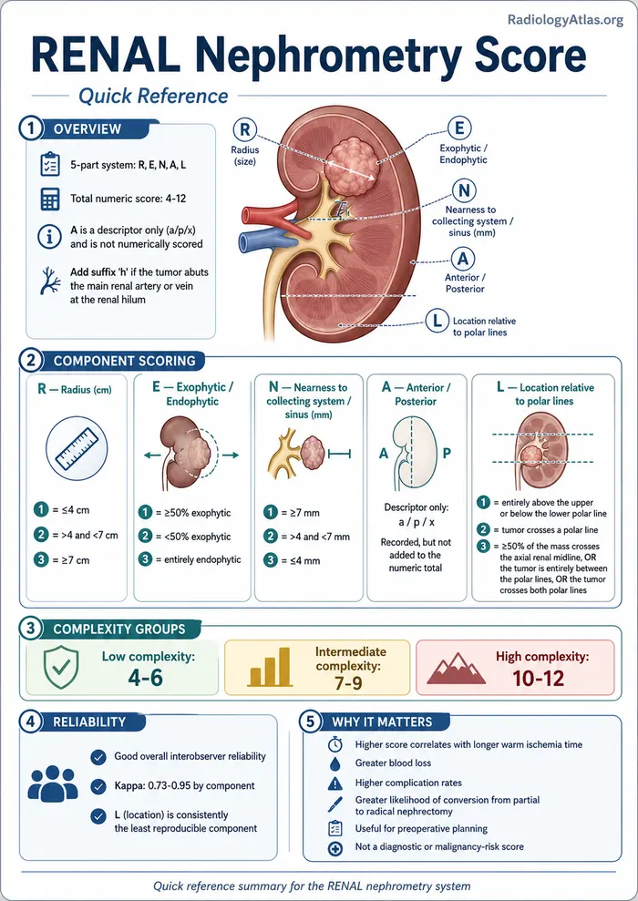
R.E.N.A.L. Nephrometry Score
The R.E.N.A.L. nephrometry score is a standardized, CT/MRI-based scoring system that quantifies the anatomic complexity of a solid renal mass using five components: Radius (maximal tumor diameter), Exophytic/endophytic properties, Nearness of the tumor to the collecting system or renal sinus, Anterior or posterior location, and Location relative to the polar lines. Each component is scored 1-3 points (with a hilar “h” suffix added when the tumor touches the main renal artery or vein), and the components are summed into a total score of 4-12 that stratifies tumors into low (4-6), intermediate (7-9), or high (10-12) complexity. Urologists use this score to counsel patients and choose between partial (nephron-sparing) and radical nephrectomy, and to anticipate operative difficulty and complication risk.
Quick Reference
- Total score range: 4-12 points, sum of 5 components (R, E, N, A, L), each scored 1-3; add suffix “h” if tumor abuts the main renal artery or vein at the renal hilum
- R (Radius, cm): 1 = ≤4 cm; 2 = >4 and <7 cm; 3 = ≥7 cm
- E (Exophytic/endophytic %): 1 = ≥50% exophytic; 2 = <50% exophytic; 3 = entirely endophytic
- N (Nearness to collecting system/sinus, mm): 1 = ≥7 mm; 2 = >4 and <7 mm; 3 = ≤4 mm
- A (Anterior/posterior): descriptor only (a/p/x), not scored numerically, but recorded and used in some outcome analyses
- L (Location relative to polar lines): 1 = entirely above the upper or below the lower polar line; 2 = tumor crosses a polar line; 3 = ≥50% of the mass crosses the axial renal midline, or the tumor is entirely between the polar lines, or the tumor crosses both polar lines
- Complexity groups: low = 4-6, intermediate = 7-9, high = 10-12
- Interobserver reliability: good overall (kappa 0.73-0.95 by component); the L (location) component is consistently the least reproducible
- Key “so what”: higher score correlates with longer warm ischemia time, greater blood loss, higher complication rates, and greater likelihood of conversion from partial to radical nephrectomy — it is a preoperative planning tool, not a diagnostic or malignancy-risk score
Background and Purpose
Kutikov and Uzzo introduced the R.E.N.A.L. nephrometry score in 2009 to give urologists and radiologists a reproducible, standardized vocabulary for describing renal tumor anatomy on cross-sectional imaging, analogous to BI-RADS in breast imaging.1,4 Before nephrometry scoring, surgical planning for renal masses relied on free-text descriptions of size and location that varied widely between readers and made it difficult to compare outcomes across surgical series. The nephrometry score converts the pertinent anatomic features of a renal mass, extracted directly from CT or MRI, into a single ordinal number that predicts operative complexity of nephron-sparing surgery.1 A related but independently derived system, the PADUA classification, was published the same year and captures overlapping anatomic domains with a different point structure.2
Imaging Anatomy: What You Are Measuring
Every component of the score is derived from the tumor’s relationship to fixed anatomic landmarks of the kidney, so accurate scoring depends on correctly identifying these structures on the source images, not just eyeballing the mass.
- Polar lines — imaginary transverse (axial) lines drawn at the level of the upper and lower margins of the renal pelvis, projected onto coronal or sagittal reformats. These lines divide the kidney into upper pole, midpole (interpolar), and lower pole segments and are the reference for the L component.
- Renal sinus fat and collecting system — the central fat-containing compartment housing the renal pelvis, infundibula, and major vessels; the tumor’s closest measured distance (in mm) to the collecting system or sinus fat, whichever is closer, defines the N component.
- Renal capsule/cortical surface — used to estimate the exophytic versus endophytic percentage of the mass (E component); a purely exophytic tumor bulges almost entirely outside the expected renal contour, while an endophytic tumor is fully contained within the normal renal contour.
- Coronal midline (anterior-posterior axis) — the tumor’s epicenter relative to a coronal plane through the kidney’s mid-portion determines the a/p/x descriptor.
- Main renal artery and vein at the hilum — direct tumor contact with these vessels triggers the hilar “h” suffix, which flags added vascular and reconstructive complexity independent of the numeric score.
Scoring Components and Criteria
| Component | 1 point | 2 points | 3 points |
|---|---|---|---|
| R — Radius (maximal diameter) | ≤4 cm | >4 cm and <7 cm | ≥7 cm |
| E — Exophytic/endophytic | ≥50% exophytic | <50% exophytic | Entirely endophytic |
| N — Nearness to collecting system/sinus | ≥7 mm | >4 mm and <7 mm | ≤4 mm |
| L — Location relative to polar lines | Entirely above upper or below lower polar line | Tumor crosses a polar line | ≥50% of mass crosses renal axial midline, tumor entirely between polar lines, or crosses both polar lines |
The A (anterior/posterior/indeterminate) descriptor is recorded as a suffix letter — a, p, or x — rather than contributing numeric points, though it has independent prognostic value in some series: an “a” or “p” descriptor (versus an indeterminate midline location) has been associated with differing complication rates in nephron-sparing surgery.5 The hilar “h” suffix is appended whenever the tumor is in direct contact with the segmental or main renal artery or vein.1,4
Total score is the sum of R + E + N + L (the A component does not add numerically): 4-6 = low complexity, 7-9 = intermediate complexity, 10-12 = high complexity.1
Imaging Technique
The nephrometry score is derived from contrast-enhanced multiphase CT (typically corticomedullary and nephrographic or excretory phases) or contrast-enhanced MRI, with thin-section axial acquisition and coronal/sagittal reformats essential for accurately locating the polar lines and measuring the tumor-to-collecting-system distance.1,4 Multiplanar reformats are not optional for reproducible scoring — axial-only interpretation systematically degrades accuracy of the L component in particular, since polar line position is best appreciated in the coronal plane.4 Measurements of tumor diameter and distance to the sinus/collecting system should be taken on the phase and plane that best displays the tumor-parenchyma and tumor-sinus interfaces, generally the corticomedullary or nephrographic phase.
Imaging Findings by Component
Radius (R): Measure the single greatest tumor diameter on whichever plane (axial, coronal, or sagittal) displays it best; this is a straightforward caliper measurement but should be performed on the phase with the sharpest tumor margin, usually corticomedullary or nephrographic phase CT or T1 post-contrast MRI.
Exophytic/endophytic (E): Estimate the percentage of tumor volume projecting beyond the expected smooth renal contour versus the percentage contained within the parenchyma. This is a visual estimate rather than a precise volumetric calculation in routine practice — round to the nearest anchor point (≥50% exophytic, <50% exophytic, or entirely endophytic/no visible cortical bulge).
Nearness to collecting system or sinus (N): On axial or coronal images, measure the shortest straight-line distance from the tumor edge to the nearest calyx, infundibulum, renal pelvis, or sinus fat margin. A tumor that abuts or invades the collecting system scores 3 regardless of overall size, reflecting the added risk of urine leak and need for collecting-system repair during resection.
Anterior/posterior (A): Assess the tumor epicenter relative to a coronal plane bisecting the kidney. Predominantly anterior tumors are more accessible via a standard laparoscopic or robotic anterior approach; predominantly posterior tumors may require repositioning or a retroperitoneal approach. Tumors that straddle the midline are recorded as “x” (indeterminate).
Location relative to polar lines (L): Identify the axial planes of the upper and lower margins of the renal pelvis on coronal reformats, then determine whether the tumor lies entirely outside these lines (polar tumor, generally more surgically favorable), crosses one polar line, or straddles the interpolar region/crosses both lines (least favorable, often closest to the hilum and collecting system).
Hilar contact (h suffix): Look specifically for tumor abutment or encasement of the segmental or main renal artery/vein at the hilum on contrast-enhanced arterial and venous phase images; this finding should be explicitly stated in the report because it changes vascular control strategy regardless of the numeric score.
Clinical Significance in Nephron-Sparing Surgery Decisions
The nephrometry score was designed specifically to inform the choice between partial nephrectomy (nephron-sparing surgery) and radical nephrectomy, and to set expectations for operative difficulty once partial nephrectomy is chosen.1 Multiple validation studies have linked higher total scores to longer warm ischemia time, greater estimated blood loss, longer operative time, and higher rates of major postoperative complications during partial nephrectomy.3,5,6,8 A UK cohort study found that each one-point increase in the RENAL score increased the odds of a higher Clavien-Dindo complication grade, and that a posterior tumor location (the “p” suffix) carried more than twice the odds of a serious complication.6 High-complexity tumors (score 10-12) are also more likely to require conversion from a planned partial nephrectomy to radical nephrectomy intraoperatively.1
The score has also been validated outside surgical resection: in patients undergoing percutaneous cryoablation or radiofrequency ablation of renal tumors, higher nephrometry scores were independently associated with both local treatment failure and major post-ablation complications, making the score useful for interventional radiology treatment planning as well as surgical planning.7 Because location relative to the collecting system and hilum drives much of this risk, the score functions less as a predictor of tumor biology and more as a map of technical difficulty — a high score does not imply a more aggressive cancer, only a more anatomically demanding resection or ablation.
Comparison with Other Nephrometry Systems
The PADUA classification, published contemporaneously, scores seven variables (including exophytic rate, renal rim location, renal sinus involvement, and tumor size) and has shown comparable predictive performance to RENAL for perioperative outcomes and conversion to radical nephrectomy.2 A comprehensive comparison of RENAL, PADUA, the NePhRO score, and the C-index found that RENAL, PADUA, and NePhRO performed comparably as independent predictors of severe complications and were each superior to the more complex C-index, which showed no significant correlation with complications in that cohort.8 In practice, RENAL remains the most widely used system in North American radiology and urology literature, while PADUA is more commonly used in European series; radiologists should be prepared to calculate either depending on institutional preference and referring surgeon.
Reproducibility and Reporting Pitfalls
Interobserver reliability of the RENAL score is generally good but uneven across components. In the original reliability study, kappa values were 0.95 for R, 0.86 for E, 0.76 for N, 0.84 for A, and only 0.73 for L — the location component was the least reproducible, with only 54% raw concordance among three observers.3 A subsequent comparison between a urologist and a radiologist scoring the same CT scans found even lower agreement for the E, A, and L components (kappa 0.47, 0.28, and 0.21, respectively), despite good agreement on R and a similar mean total score between readers, underscoring that the total score can converge even when individual component scoring diverges.5 Common sources of disagreement include failing to use coronal reformats to localize the polar lines, inconsistent rounding of exophytic/endophytic percentage at the 50% boundary, and disagreement about what counts as “touching” the collecting system versus a few millimeters of separation. Radiologists reporting a nephrometry score should explicitly state the total score, each component letter-value, and the hilar suffix if present, rather than a single number alone, so the referring surgeon can see which specific anatomic feature is driving the complexity.
Differential Considerations and Practical Notes
The nephrometry score is not a diagnostic tool and does not by itself distinguish renal cell carcinoma from a benign mass such as oncocytoma or fat-poor angiomyolipoma — it should be applied to any solid, indeterminate, or known-malignant renal mass being considered for nephron-sparing management, after the mass has already been characterized on standard renal mass CT/MRI protocol. Cystic renal masses are typically excluded from or scored separately under nephrometry systems, as the underlying scoring criteria were validated on solid tumors. Because polar line position and sinus fat distance can be difficult to judge on limited or motion-degraded studies, a dedicated renal mass protocol with thin sections and multiplanar reformats should be requested when a nephrometry score is specifically needed for surgical planning, rather than deriving it from a suboptimal single-phase abdominal CT.
FAQ
What does a R.E.N.A.L. nephrometry score of 10 or higher mean?
A score of 10-12 places a renal tumor in the high-complexity category, which is associated with longer warm ischemia time, greater blood loss, higher rates of major complications, and a greater chance of conversion from partial to radical nephrectomy during surgery.1,6
Who calculates the R.E.N.A.L. nephrometry score, the radiologist or the urologist?
Either can calculate it from the same CT or MRI, but studies show only moderate agreement between radiologists and urologists on individual components (especially exophytic/endophytic percentage, anterior/posterior location, and polar line location), even though total scores and downstream complication associations tend to be similar between readers.5 Many practices now have radiologists report the full score as part of the renal mass CT/MRI interpretation.
Is the R.E.N.A.L. nephrometry score used to decide if a mass is cancerous?
No. The score describes anatomic complexity and surgical difficulty, not malignant potential; a low-complexity score does not mean a mass is benign, and a high-complexity score does not mean it is more aggressive cancer.1
What is the difference between the RENAL score and the PADUA score?
Both are CT/MRI-based anatomic scoring systems for renal masses developed around the same time; PADUA uses seven variables and a different point scale, but head-to-head comparisons show RENAL and PADUA have similar accuracy for predicting perioperative complications and conversion to radical nephrectomy.2,8
Which nephrometry component is hardest to score consistently?
The L component (tumor location relative to the polar lines) is consistently the least reproducible across validation studies, due to variability in identifying the polar lines and in judging how much of the tumor crosses them.3,5
Does the R.E.N.A.L. score apply to tumors treated with ablation instead of surgery?
Yes. In a series of 751 renal tumors treated with percutaneous cryoablation or radiofrequency ablation, higher RENAL nephrometry scores were significantly associated with both local treatment failure and major complications, so the score is also used for interventional radiology treatment planning.7
References
- Kutikov A, Uzzo RG. The R.E.N.A.L. nephrometry score: a comprehensive standardized system for quantitating renal tumor size, location and depth. J Urol. 2009;182(3):844-853. PMID: 19616235
- Ficarra V, Novara G, Secco S, et al. Preoperative aspects and dimensions used for an anatomical (PADUA) classification of renal tumours in patients who are candidates for nephron-sparing surgery. Eur Urol. 2009;56(5):786-793. PMID: 19665284
- Kolla SB, Spiess PE, Sexton WJ. Interobserver reliability of the RENAL nephrometry scoring system. Urology. 2011;78(3):592-594. PMID: 21782219
- Parsons RB, Canter D, Kutikov A, Uzzo RG. RENAL nephrometry scoring system: the radiologist’s perspective. AJR Am J Roentgenol. 2012;199(3):W355-W359. PMID: 22915426
- Benadiba S, Verin AL, Pignot G, et al. Are urologists and radiologists equally effective in determining the RENAL Nephrometry score? Ann Surg Oncol. 2015;22(5):1618-1624. PMID: 25384701
- Reddy UD, Pillai R, Parker RA, et al. Prediction of complications after partial nephrectomy by RENAL nephrometry score. Ann R Coll Surg Engl. 2014;96(6):475-479. PMID: 25198982
- Schmit GD, Thompson RH, Kurup AN, et al. Usefulness of R.E.N.A.L. nephrometry scoring system for predicting outcomes and complications of percutaneous ablation of 751 renal tumors. J Urol. 2013;189(1):30-35. PMID: 23164375
- Kriegmair MC, Mandel P, Moses A, et al. Defining renal masses: comprehensive comparison of RENAL, PADUA, NePhRO, and C-index score. Clin Genitourin Cancer. 2017;15(2):248-255. PMID: 27594556