
TI-RADS (ACR Thyroid Imaging Reporting and Data System)
 sakthi379
 TI-RADS (ACR Thyroid Imaging Reporting and Data System)
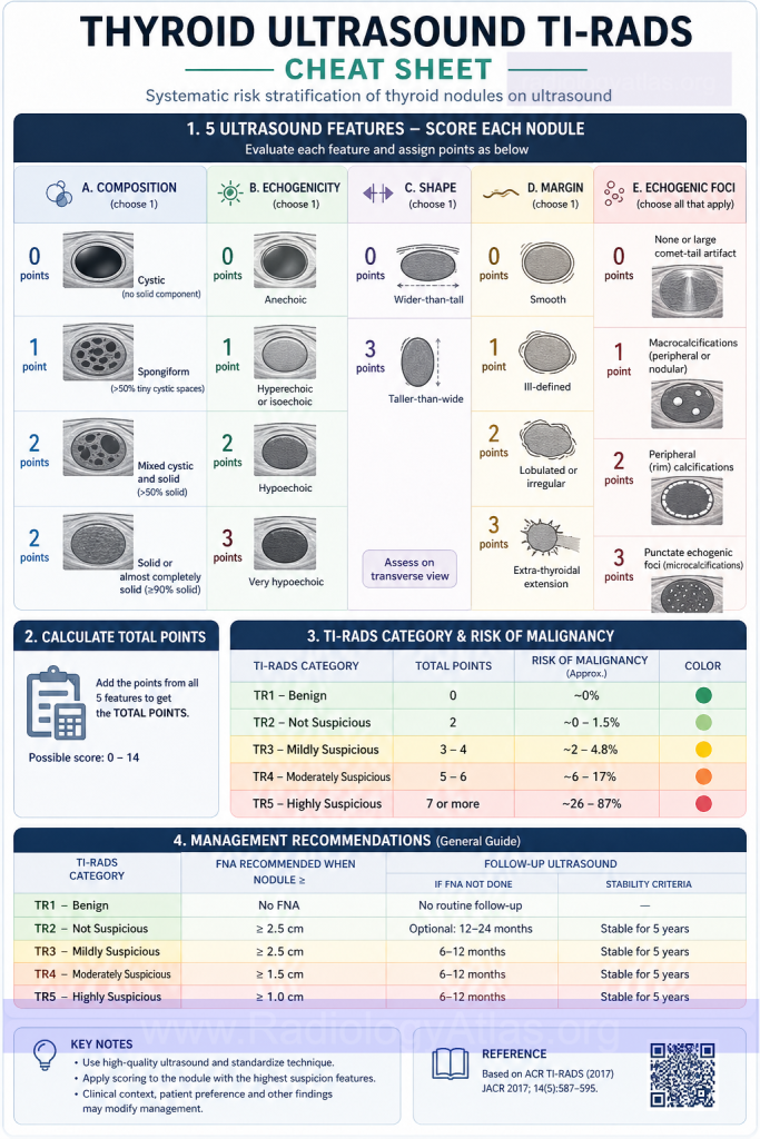
ACR TI-RADS is a point-based ultrasound risk-stratification system for thyroid nodules that sums scores across five sonographic categories — composition, echogenicity, shape, margin, and echogenic foci — to assign a nodule to one of five risk tiers, TR1 (benign) through TR5 (highly suspicious). The tier and the nodule’s maximum diameter together determine whether the recommendation is no follow-up, ultrasound surveillance, or fine-needle aspiration (FNA).
Quick Reference
- Five feature categories, points summed: Composition (0–2), Echogenicity (0–3), Shape (0/3), Margin (0–3), Echogenic Foci (0–3, additive)
- TR1 (0 pts): Benign — no FNA
- TR2 (2 pts): Not suspicious — no FNA
- TR3 (3 pts): Mildly suspicious — FNA if ≥2.5 cm, follow if ≥1.5 cm
- TR4 (4–6 pts): Moderately suspicious — FNA if ≥1.5 cm, follow if ≥1.0 cm
- TR5 (≥7 pts): Highly suspicious — FNA if ≥1.0 cm, follow if ≥0.5 cm
- “Taller-than-wide” shape is the only feature that alone scores 3 points and can push a nodule to TR4/TR5
- Punctate echogenic foci (suspicious for microcalcifications) score 3 points; peripheral/rim calcifications score 2; macrocalcifications score 1
- Purely cystic nodules are TR1 by definition and never require FNA
- So what: Higher TI-RADS category correlates with higher malignancy rate on cytology, and applying size cutoffs by category reduces unnecessary biopsies without a large increase in missed cancers
Interactive TI-RADS Calculator
Open calculator — toggle features and diameter to get score, TR category, and recommendation
Toggle the sonographic features seen on your image and enter the nodule’s maximum diameter to get the composite score, TR category, and management recommendation.
TR1
Educational reference tool based on ACR TI-RADS (Tessler et al, 2017). Does not replace clinical judgment or a formal radiology report.
Background
Thyroid nodules are extremely common on cervical ultrasound, and most are benign. Before structured risk stratification, biopsy decisions relied heavily on size and gestalt, which drove a high volume of FNAs for nodules with a low pretest probability of malignancy. The ACR TI-RADS Committee published its white paper in 2017, adapting the BI-RADS/lexicon concept from breast imaging to thyroid ultrasound: standardized descriptors, a points-based composite score, and size-based action thresholds tied to risk category (Tessler et al., 2017). The goal was to reduce interobserver variability in nodule description and to limit biopsy to nodules whose sonographic appearance actually predicts malignancy risk.
Imaging Anatomy
TI-RADS is assessed entirely on grayscale (± color Doppler, which is not scored) cervical ultrasound of the thyroid gland. Relevant anatomy for characterization includes:
- Thyroid parenchyma — used as the internal reference for echogenicity comparison (nodule echogenicity is graded relative to the adjacent normal gland, not to strap muscle)
- Nodule margin and capsule interface — the border between the nodule and surrounding parenchyma, evaluated for smoothness, irregularity, lobulation, and extrathyroidal extension
- Long axis relative to the skin surface — the anteroposterior-to-transverse ratio on a transverse image defines “taller-than-wide” shape, best assessed on the image where the nodule appears roundest
- Internal architecture — solid vs. cystic vs. mixed components, and the distribution of any echogenic foci within the nodule
- Strap muscles and trachea — landmarks used to judge extrathyroidal extension and to orient transverse vs. sagittal planes
Imaging Findings and Scoring
Each nodule is scored feature-by-feature on grayscale ultrasound; points from each category are added for a composite score that maps to a TR level.
Composition (choose one, 0–2 points)
| Finding | Points |
|---|---|
| Cystic or almost completely cystic | 0 |
| Spongiform | 0 |
| Mixed cystic and solid | 1 |
| Solid or almost completely solid | 2 |
Echogenicity (choose one, relative to thyroid parenchyma, 0–3 points)
| Finding | Points |
|---|---|
| Anechoic | 0 |
| Hyperechoic or isoechoic | 1 |
| Hypoechoic | 2 |
| Very hypoechoic (more hypoechoic than strap muscle) | 3 |
Shape (choose one, 0 or 3 points)
Taller-than-wide on a transverse image scores 3 points; wider-than-tall scores 0. This is assessed independent of composition or echogenicity and is the single most efficient feature for flagging malignancy risk when present.
Margin (choose one, 0–3 points)
| Finding | Points |
|---|---|
| Smooth | 0 |
| Ill-defined | 0 |
| Lobulated or irregular | 2 |
| Extrathyroidal extension | 3 |
Echogenic Foci (select all that apply, additive, 0–3 points)
| Finding | Points |
|---|---|
| None or large comet-tail artifacts | 0 |
| Macrocalcifications | 1 |
| Peripheral (rim) calcifications | 2 |
| Punctate echogenic foci (suspicious for microcalcifications) | 3 |
Composite Score to TR Category
| Total points | Category | Assessment |
|---|---|---|
| 0 | TR1 | Benign |
| 2 | TR2 | Not suspicious |
| 3 | TR3 | Mildly suspicious |
| 4–6 | TR4 | Moderately suspicious |
| ≥7 | TR5 | Highly suspicious |
Multi-institutional validation of this scheme found that malignancy rates rise stepwise from TR1/TR2 through TR5, supporting the size-stratified FNA thresholds (Middleton et al., 2017; Grant et al., 2018).
Management Thresholds by Category
| Category | FNA if diameter ≥ | Follow-up if diameter ≥ |
|---|---|---|
| TR1 (Benign) | No FNA | No follow-up |
| TR2 (Not suspicious) | No FNA | No follow-up |
| TR3 (Mildly suspicious) | 2.5 cm | 1.5 cm |
| TR4 (Moderately suspicious) | 1.5 cm | 1.0 cm |
| TR5 (Highly suspicious) | 1.0 cm | 0.5 cm |
Differential Considerations and Pitfalls
- Spongiform nodules (multiple microcystic spaces occupying >50% of nodule volume) score 0 for composition and behave as benign even if other features are equivocal — don’t over-call these based on size alone.
- Comet-tail artifacts within cystic components (colloid) are a benign echogenic focus and score 0; they are frequently mistaken for punctate calcifications, which score the maximum 3 points — distinguishing the two on real-time scanning is essential.
- Echogenicity must be compared to adjacent normal thyroid parenchyma, not to strap muscle, except when specifically grading “very hypoechoic” (more hypoechoic than strap muscle).
- Taller-than-wide shape should be assessed on the transverse image at the point where the nodule looks roundest; measuring on an oblique plane can produce a false-positive or false-negative shape call.
- Partially cystic nodules should have both the solid component’s margin and echogenic foci graded on the solid portion, not the cystic component.
- ACR TI-RADS was developed and validated primarily in adults; pediatric thyroid nodules and nodules in patients with a history of medullary thyroid carcinoma or hereditary cancer syndromes may not follow the same risk gradient, and clinical context should modify management even when the sonographic score is reassuring.
Clinical Impact
TI-RADS reporting standardizes nodule description in the impression (composition, echogenicity, shape, margin, echogenic foci, composite score, and TR category), which lets referring clinicians and downstream readers apply consistent, size-adjusted FNA thresholds rather than reflexively biopsying by size alone. Retrospective cohort data show that applying ACR TI-RADS criteria is specific for identifying benign nodules and substantially limits the number of FNAs performed, while missing only a small proportion of malignancies, and that higher composite scores correlate with a stepwise increase in malignant cytology on FNA (Koseoglu Atilla et al., 2018; Modi et al., 2020). Category should be stated explicitly in the report impression along with the specific recommendation (no follow-up, ultrasound surveillance interval, or FNA) so the referring provider does not have to re-derive the score from the description.
Frequently Asked Questions
What does a TI-RADS score of TR4 mean?
TR4 (“moderately suspicious”) corresponds to a composite score of 4–6 points across the five ACR TI-RADS categories. FNA is recommended for TR4 nodules measuring 1.5 cm or larger, and ultrasound follow-up is recommended for those 1.0 cm or larger.
What ultrasound features are scored in ACR TI-RADS?
Five features are scored: composition (cystic to solid), echogenicity (relative to thyroid parenchyma), shape (taller-than-wide vs. wider-than-tall), margin (smooth to extrathyroidal extension), and echogenic foci (none/comet-tail to punctate echogenic foci). Points from each category sum to a composite score that determines the TR category.
Do all TI-RADS categories require biopsy?
No. TR1 and TR2 nodules never require FNA regardless of size. TR3–TR5 nodules require FNA only once they reach a category-specific size threshold — 2.5 cm for TR3, 1.5 cm for TR4, and 1.0 cm for TR5.
What is a “taller-than-wide” nodule and why does it matter?
A taller-than-wide nodule has an anteroposterior dimension greater than its transverse dimension on a transverse ultrasound image. It is the only shape finding that scores points (3 points if present) and reflects growth that crosses normal tissue planes, a feature associated with malignancy even when other features are unremarkable.
Are punctate echogenic foci the same as microcalcifications?
Punctate echogenic foci are the sonographic correlate suspected to represent microcalcifications (often psammoma bodies in papillary carcinoma), but pathologic confirmation is required to call them true microcalcifications. On ACR TI-RADS they carry the maximum 3 points in the echogenic foci category and should be distinguished from benign comet-tail artifacts arising from colloid.
How does ACR TI-RADS compare with other thyroid risk-stratification systems?
ACR TI-RADS is one of several point- or pattern-based systems (others include ATA guidelines, Kwak TI-RADS, and EU-TIRADS). Comparative studies show broadly similar diagnostic performance for malignancy detection across systems, with ACR TI-RADS’s additive, size-adjusted structure aimed particularly at minimizing unnecessary FNA of low-risk nodules (Migda et al., 2023).
References
- Tessler FN, Middleton WD, Grant EG, et al. ACR Thyroid Imaging, Reporting and Data System (TI-RADS): White Paper of the ACR TI-RADS Committee. J Am Coll Radiol. 2017;14(5):587-595. PMID: 28372962
- Middleton WD, Teefey SA, Reading CC, et al. Multiinstitutional Analysis of Thyroid Nodule Risk Stratification Using the American College of Radiology Thyroid Imaging Reporting and Data System. AJR Am J Roentgenol. 2017;208(6):1331-1341. PMID: 28402167
- Grant EG, Tessler FN, Hoang JK, et al. Thyroid Imaging Reporting and Data System (TI-RADS): A User’s Guide. Radiology. 2018;287(2):355-359. PMID: 29558300
- Koseoglu Atilla FD, Ozgen Saydam B, Erarslan NA, Diniz Unlu AG, Yilmaz Yasar H, Ozer M, Akinci B. Does the ACR TI-RADS scoring allow us to safely avoid unnecessary thyroid biopsy? single center analysis in a large cohort. Endocrine. 2018;61(3):398-402. PMID: 29744655
- Modi L, Sun W, Shafizadeh N, Negron R, Yee-Chang M, Zhou F, Simsir A, Sheth S, Brandler TC. Does a higher American College of Radiology Thyroid Imaging Reporting and Data System (ACR TI-RADS) score forecast an increased risk of malignancy? A correlation study of ACR TI-RADS with FNA cytology in the evaluation of thyroid nodules. Cancer Cytopathol. 2020;128(7):470-481. PMID: 32078249
- Migda B, Migda M, Migda MS, Slapa RZ. Diagnostic performance of the EU TI-RADS and ACR TI-RADS scoring systems in predicting thyroid malignancy. Wideochir Inne Tech Maloinwazyjne. 2023;18(2):303-311. PMCID: PMC10335610
- Pires AT, Mustafá AMM, Magalhães MOG. The 2017 ACR TI-RADS: pictorial essay. Radiol Bras. 2022;55(1):47-53. PMID: 35210664, PMCID: PMC8864691